Introduction
Optical Coherence Tomography (OCT) has emerged as a breakthrough technique for non-destructive three-dimensional (3D) imaging of tissue and materials. By utilizing the principles of Fourier Transform and image generation, OCT has found applications in diverse fields, including research, medical diagnostics, guided surgery, industrial processing, and non-destructive testing. This article aims to provide an in-depth understanding of OCT’s working principle, its significance in various domains, and its remarkable impact on fields like ophthalmology and neuroscience.
Working Principle of OCT
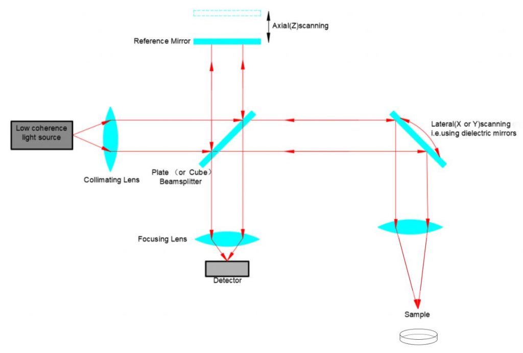
At its core, OCT is an interferometric imaging technique that relies on the interference of light waves. It uses low-coherence interferometry, where a broadband light source emits near-infrared light, typically in the range of 800 to 1,300 nm. This light is split into a sample arm and a reference arm. The sample arm directs the light towards the object or tissue under investigation, while the reference arm contains a reference mirror. The light reflected back from the sample arm and reference arm is combined, and interference between the two beams is measured.
Fourier Transform and Image Generation
To generate 3D images, OCT employs Fourier Transform techniques. The interference pattern obtained from the combined beams is processed using Fourier Transform algorithms to extract depth information. By scanning the sample arm along one axis, a series of A-scans (depth profiles) are acquired. These A-scans, when combined, form a two-dimensional (2D) cross-sectional image known as a B-scan. By sequentially acquiring multiple B-scans, a 3D volumetric image can be reconstructed.
Applications of OCT
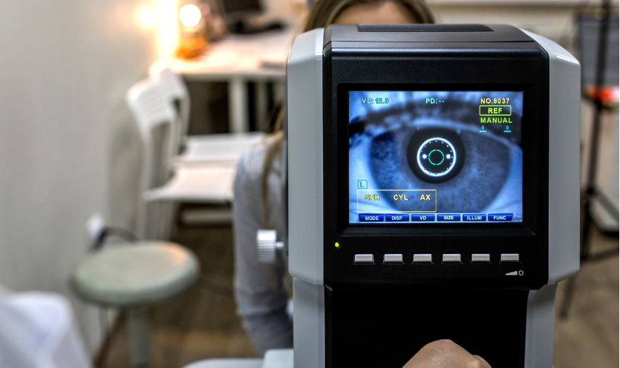
1. Ophthalmology: OCT has revolutionized the field of ophthalmology by enabling high-resolution imaging of ocular structures. It has become an indispensable tool for diagnosing and monitoring diseases such as macular degeneration, glaucoma, diabetic retinopathy, and retinal detachment. OCT’s ability to visualize retinal layers and quantify their thickness has greatly enhanced early detection and treatment planning. Additionally, advancements in asphere and micro optics have improved OCT’s imaging capabilities, allowing for even finer details to be captured.
2. Neuroscience: OCT plays a crucial role in neuroscience research by facilitating non-invasive imaging of neural tissue. It enables the visualization of brain structures and the study of neurodegenerative disorders such as multiple sclerosis and Alzheimer’s disease. OCT’s high spatial resolution and depth penetration allow researchers to examine subtle changes in neuronal morphology and track disease progression. The integration of medical optics and OCT has opened up new possibilities for studying brain function and advancing our understanding of the nervous system.

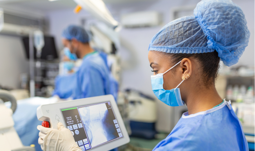
3. Guided Surgery: In surgical applications, OCT provides real-time, high-resolution imaging of tissue microstructure, aiding surgeons in making precise and informed decisions. It assists in identifying tumor margins, assessing tissue viability, and guiding the placement of surgical instruments. OCT’s ability to visualize subsurface tissue layers reduces the risk of incomplete tumor removal and improves surgical outcomes.
4. Industrial Processing and Non-Destructive Testing: OCT finds utility in industrial applications such as quality control, material inspection, and non-destructive testing. It enables real-time monitoring of manufacturing processes, detecting defects or irregularities in materials, and ensuring product quality. Additionally, OCT is employed in the evaluation of coatings, paints, and thin films, providing detailed structural information and enhancing the efficiency of industrial processes.

Conclusion
Optical Coherence Tomography has emerged as a powerful and versatile imaging technique with wide-ranging applications in research, medical diagnostics, guided surgery, industrial processing, and non-destructive testing. By harnessing the principles of interferometry, Fourier Transform, and image generation, OCT enables non-destructive 3D imaging of tissue and materials with remarkable spatial resolution and depth penetration. Its impact in fields such as ophthalmology and neuroscience has been transformative, revolutionizing disease diagnosis, treatment planning, and research methodologies. As OCT continues to advance, it holds immense potential for further advancements in diverse fields, contributing to improved healthcare outcomes, enhanced industrial processes, and expanded scientific understanding.
Avantier Inc. offers custom imaging solutions along with manufacturing services for the high quality optical components that are used in OCT instruments including dichroic mirrors, filters, beamsplitters, collimation lenses, scan lenses, objective lenses, etc. Please contact us if you’d like to schedule a free consultation or request for quote on your next project.
For other articles on OCT, please click here
Optical Coherence Tomography (OCT)
Advancements in Non-Destructive Testing : OCT Part 3