
Key Takeaways
- FLIM Advances Diagnostics: FLIM offers precise views of living cells and proteins, aiding diagnoses and enhancing understanding of diseases like cancer.
- TCSPC Measures Fluorescence Lifetimes: TCSPC captures differences in fluorescence lifetime, enabling picosecond-level measurements crucial for medical imaging.
- FRET Enhances Protein Studies: FRET microscopy, combined with FLIM, provides detailed insights into protein interactions, aiding biomedical research and diagnostics.
Fluorescence-Lifetime Imaging
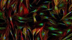
One powerful technique used in medical diagnostics research is fluorescence-lifetime-imaging (FLIM). Precision lasers, fluorescent dyes, and state of the art recording devices enable medical professionals and researchers to view living cells, sub-cell interactions, and the proteins responsible for them with accuracy and precision. This makes FLIM invaluable for clinical diagnosis and also provides an entirely new, molecular-level understanding of immune responses, allergies, and even cancer.
But what is really happening when you generate an image using FLIM? The key information your image is based on is actually the differences in the fluorescence lifetime of excited fluorophore dyes, which may be used to tag different proteins or parts of proteins.
At its simplest, one can think of fluorescence lifetime microscopy as a laser beam scanning across a sample that is placed under a microscope. Fluorescence can be measured at every position across the sample. The rate at which the fluorescence intensity decreases depends on the pH, ion concentration, and fluorophore concentration of the biochemical sample as well as on the refractive index of the environment. Differences in the lifetime of fluorescence signals difference in environment.
What is Time-Correlated Single-Photon Counting?
Most fluorescence lifetime imaging today is done by time-correlated single-photon counting (TCSPC). TCSPC enables us to quantify the time between an excitation pulse (delivered by laser) and one emitted photon. The arrival of the photon is measured by a sensitive detector which produces a separate electric pulse at each arrival. This detector may be a single photon avalanche photo diode (SPAD) or a photo-multiplier tube (PMT).
When a large number of times have been recorded, the photon counts can be sorted by arrival time into an intensity-based histogram. Modern methods enable millions of start-stop times to be recorded over a reasonably sized interval.

What is the Gating Method?
An alternative method of conducting FLIM is the gating method. In this method part of the laser pulse, en route to the sample, is reflected by a dichroic mirror onto a photodiode. This photodiode activates a delay generator that controls a gated optical intensifier (GOI) which is placed in front of a CCD (charge-coupled device) detector. Each time the GOI is opened, it allows detection for an extremely short time interval. When fluorescence emission is collected after multiple delay times, this system can record a comprehensive view of the time range of the fluorescence decay of the sample. Modern integrated intensified CCD cameras allow sub-nano-second resolution FLIM.
Fluorescence Resonance Energy Transfer (FRET) Microscopy
FRET microscopy allows us to view protein-protein interactions closer up than is possible with FLIM alone. When two fluorophores are less than 10 nanometers apart excitation can be transferred between them without photons. This transfer is distance-dependent, with the efficiency increasing at a rate proportional to the inverse sixth power of the separation between the molecules. FRET is often combined with FLIM to determine the exact spatial proximity of proteins and molecules.
Biomedical Imaging and Diagnostics
Although conventional imaging techniques have been key to many medical diagnoses since the birth of modern medicine, they are limited in the information they can provide about biological processes and the onset of diseases. A better understanding of pathological and normal tissue composition, morphology, and function is dependent on better imaging techniques that allow us to see biological processes in situ.
Imaging techniques for biomedical applications is a quickly growing field, and we’ve seen some enormous advances in recent years. Advances in fluorescent lifetime imaging, for instance, have transformed many areas of medical diagnostics. A combination of high lasers, versatile beam scanners, sensitive detectors and high power cameras enable researchers to view biological tissue in vivo and gain valuable insights into both disease and healthy physiology.
Optical Imaging Techniques: Viewing Tissue at Depth
Biological tissue is turbid and inherently difficult to image because of its dense, inhomogeneous nature. Tissue scattering has made deep tissue imaging highly problematic in the past. But two-photon fluorescence microscopy has been successful at producing high resolution imaging even at slightly deeper tissue levels, and today new developments are enabling high-quality imaging at a deeper level than ever before.
Avantier Inc is at the forefront of imaging systems solutions and optical solutions, and we are proud of our track record of providing researchers, medical practitioners and industry clients the optics they need to push the boundaries with high-performance imaging. Contact us for more information on how we can partner with you.


")